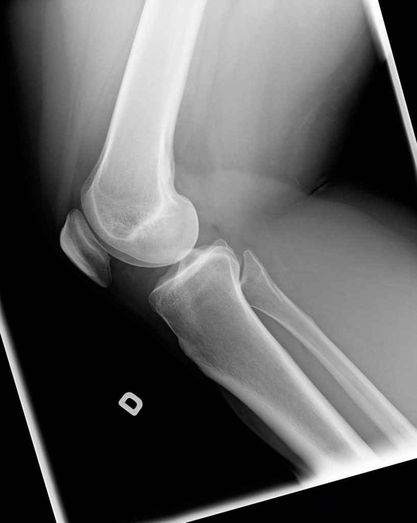
Patella or kneecap dislocation refers to the abnormal sideways movement of the patella. This is a movement out of its normal position. The normal movement of the patella is up and down but when it dislocates, it will move sideways.
The patella is a triangular bone located at the front of the knee to which the quadriceps tendon attaches.

In this article, we shall talk about what causes the patella to dislocate and how knee cap dislocation is treated.
The patella commonly dislocates to the outside of the knee (lateral side ) rather than the inside of the knee (medial side).
Difference between a patella dislocation and a patella subluxation
When the patella subluxates, it moves over to the edge of the knee joint. In a way, a subluxation is like an incomplete dislocation.
A patella dislocation on the other hand means that the patella completely moved out of the joint and can be seen on the side.
Patella dislocation and patella subluxation make up the spectrum known as patella instability.
Causes of patella dislocations
- Trauma. The most common cause of dislocation is traumatic.
That being said, there are risk factors that put one at a higher risk of dislocating. These are;
- Knock knee alignment. This is medically termed genu valgum.
- Female gender; there is a higher incidence of patella dislocation in women than in men. This could be because women may be more flexible than men.
- Family history of patella dislocations. You are more likely to dislocate your kneecap if someone in your family like a biological parent or first-degree relative has it.
- Hyperlaxity of the joint. This means having joints that are too stretchy. People with hyper lax joints are more likely to have a patella dislocation than their counterparts.
There are some conditions characterised with hyperlaxity and these include; Ehler Danlos syndrome, Marfan’s syndrome, Down’s syndrome.
- Anatomical variation at the knee. This includes features like a lateral tibial tubercle, a high-riding patella, shallow trochlear groove.
Signs and symptoms of patella dislocation
- Pop in the knee. You will hear a loud pop at the kneee when the patella dislocates. This is common in first-time dislocations.
- Knee pain. The pain is worse during the first dislocation. It may continue to be painful every time the patella dislocates but not as bad as the 1st time.
- Swelling of the knee
- Failure to straighten the leg
- Inability to walk on that leg
- Apprehension. You may feel apprehensive when someone touches your knees because of fear that they will dislocate your patella. Dislocation of a joint is an uncomfortable experience.
- Wasting of the quadriceps; This is more noticeable if only one side dislocates and will be noticed after a week or more of patella dislocation. The patella is a vital part of the extensor mechanism.
The extensor mechanism comprises the quadriceps muscles, the patella, and the patella tendon. Therefore its absence does affect the overall function of the quadriceps mechanism.
At your doctor’s, there are some things that the doctor will look out for;
- Positive J sign.
A positive J sign is seen when the cap translates out of its normal groove when the knee is straightened from a flexed position but will move back to its position when nearing full extension of the leg.
-Since patella instability can be present in both knees, your doctor will often assess both knees during your visit.
Types of patella dislocations
1. Acute patella dislocations
It’s called that if it is the first time it has happened. This often is a result of trauma.
2. Recurrent dislocation
Any dislocation after the acute event is termed recurrent
3. Habitual dislocation
Habitual dislocations occur with any flexion or bending of the knee
Investigations for patella dislocation
Most kneecaps relocate to the normal position before you get to the doctor’s office but routinely, you will need;
- X-rays.
Why are X-rays taken
- To rule out a fracture
Sometimes, you may have a fracture of your patella or femur associated with the dislocation. This may be picked on the x-ray.
- It checks for the alignment of your legs.
In case you have knock knees or bowed knees, the doctor may do alignment X-rays as this may need to be corrected if there is any surgical correction needed.
2. Ct scans
CT scans are done to check the rotational profile of the leg to be operated
3. MRI scan
In the event of fractures that involve the cartilage, an MRI is needed as these will not be seen easily by other radiological modalities.
Also, an MRI will show if there are other ligaments injured inside your knee.
Treatment of patella dislocation
Initial treatment of patella dislocation
Initial treatment of patella dislocation
- Pain management
A patella dislocation is very painful so in the emergency unit, the doctor or nurse will give you pain medication
- Reduction of the patella dislocation
This will normally be done with you sleeping or sedated and relaxed. The doctor will simply pop the knee cap back to its proper location and then place you in a brace.
In some cases, you may get to the hospital when the patella has already fallen back into place. In that case, the doctor will just place you into a brace.
Non-operative treatment
- Physiotherapy
Quadriceps strengthening exercises;
Vastus medialis obliqus or VMO strengthening exercises. The small muscle that attaches close to the inner aspect of the knee needs strengthening. Your doctor will give you special exercises and send you to a physiotherapist.
- Bracing.
Braces are used as static stabilizers that prevent the knee cap from moving out of the normal position.
Surgery for patella dislocation
- Young children.
A young child is one who still has growth plates.
In children who still have growth plates, all surgeries are done away from the growth plate to prevent injury to the growth plates. The surgeries are often soft tissue procedures.
Soft tissue procedures can involve reconstructions or repair of ligaments and tendons and release of tight structures to prevent redislocation
Some of these surgeries include;
- MPFL (medial patellofemoral ligament) reconstruction.
In this surgery, the surgeon will create a new ligament that is attached from the inner and lower aspect of your femur connecting to the inner side of the patella. The tendon used to create your new ligament can either be yours (autograft) or can be donated (allograft).
2. Lateral release; A lateral release is done by releasing the tight structures on the outer side of the knee. This is commonly the lateral retinacular but can also include the capsules of the joint.
3. VMO advancement. VMO is short for vastus medialis obliquus. In this procedure, the attachment of the muscle is moved to give more of an inward pull to the patella.
4. Guided growth surgery.
Guided growth surgery is done for children with open growth plates (physes) by placement of plates and screw adjacent to the growth plates to correct either knock knees or bowed legs.
- Older children
Together with repair or reconstruction of soft tissue procedures, surgeries involving cutting and repositioning of bones can be done because the growth plates have closed.
- Tibial tubercle osteotomy (TTO).
The tibial tubercle is the small tiny bump on the shin just below your knee joint. In a TTO, it will be transferred more inward and moved slightly lower. It will be fixed in place with screws which can later be taken out after the tibial tubercle heals.
Recovery from patella dislocation surgery
The recovery time depends on what is done. Soft tissue surgeries heal faster
- Bracing or cast after surgery
The cast or Brace provides immobilization and protection to the knee during the early weeks of recovery.
- Non-weight bearing after surgery
The time you will stay off your feet following surgery depends on the kind of surgery done. It often varies from 2 weeks to 6 weeks.
Prevention of patella dislocation
In the presence of the risk factors that we mentioned above, it could be hard to 100% prevent patella dislocations but that being said, rehabilitation and strengthening of the quadriceps is a good place to start.
Prognosis after patella dislocation;
After the first dislocation, there is a 40% risk of dislocation. The percentage goes higher after that and it’s almost at 100% after the 3rd dislocation.
What to expect after surgery for patella dislocation
0- 2 weeks This is a time to let the wound heal and get the post-op swelling to settle down.
You are not allowed to walk on your leg.
You will probably be in a brace or a cast to keep your knee in an extended position.
2 to 6 weeks You will continue to be in a cast or brace and will not be allowed to walk on that leg.
6 to 8 weeks You may start to gently move your knee. If the X-rays show signs of union in the bones, your doctor may allow you to gently start putting weight on that leg.
8 weeks to 3 months You could start physiotherapy to rehabilitate your quadriceps and improve your knee range of motion.
In conclusion, patella dislocations are quite common, more in females than males, and can be crippling in some cases. There is a higher risk of redislocation with every increasing episode of dislocation.
Treatment can be either nonsurgical or surgical and this is guided by the frequency of the dislocations and your age.
