Microfracture surgery refers to the process of surgically creating small blunt holes in the bone underneath the damaged cartilage to stimulate its repair by formation of a new kind of cartilage known as fibrocartilage.
The cartilage found in joints is a very unique biological structure. Once this kind of cartilage is injured, it is unable to recover with the same exact composition like it was originally.
In this article, we will discuss how microfracture of cartilage works and also elaborate on the different physiotherapy done for recovery.
Injured cartilage heals with a mixture of cartilage and fibrous tissue which is known as fibrocartilage. This concept of creating fibrocartilage is applied in the treatment of defects in the cartilage particularly in the knee joint.
The use of microfracture surgery has been reported to be effective in improving the quality of life of patients in regard to pain relief and better function.
When is microfracture surgery done for children?
Osteochondritis dissecans also known as OCDs is the main reason for doing microfracture surgery in children.
Osteochondritis dissecans happens when there is death of the articular cartilage due to lack of blood or nutrients reaching the articular cartilage.
Osteochondritis dissecans follows repetitive trauma to a joint and is most common in weight-bearing joints.
Most affected joints by Osteochondritis dissecans
- Knee joint.
The lower part of the femur is the most affected. This is the most common site of OCDs.
- Elbow joint
The capitellum is a bone on the lower end of the humerus which is most affected in gymnasts.
-ankle joint
The talus is a bone in the foot that makes up the ankle joint and is also a common site for OCDs
The surgical procedure of microfracture surgery
This surgery is done by an orthopedic surgeon. You will require anesthesia for this operation.
The orthopedic surgeon will make small incisions on the skin which will measure about 0.5cm and through these, access the inside of your knee joint.
During arthroscopic surgery which is the kind where a mini-camera is used to visualize inside your knee joint, areas of degenerative cartilage can be chipped away to form small holes which eventually fill up with fibrocartilage.
In this surgical procedure, small holes are made in the subchondral bone (bone underneath the cartilage) using surgical equipment.
Blood and fat released from the drilled bone will form a mesenchymal clot which is rich in factors that lead to the formation of a new type of cartilage.
Sometimes, a larger piece of cartilage is detached from the bone. If that piece of cartilage is still attached to the bone in some areas or still has bone on it’s undersurface, it will be fixed back.
If the fragment of cartilage is freely floating around the joint, then it is removed because it can cause irritation and blockage of the joint.
At the end of the surgery, these small wounds made on the skin will be repaired with sutures.
What happens after the surgery
This is normally a day surgery and you will be discharged home the same day after the surgery.
You will be given an appointment to see your doctor in 10 to 14 days to assess the wounds.
You will continue to use crutches and won’t be allowed to step on that leg for about 6 to 8 weeks from the day of the surgery.
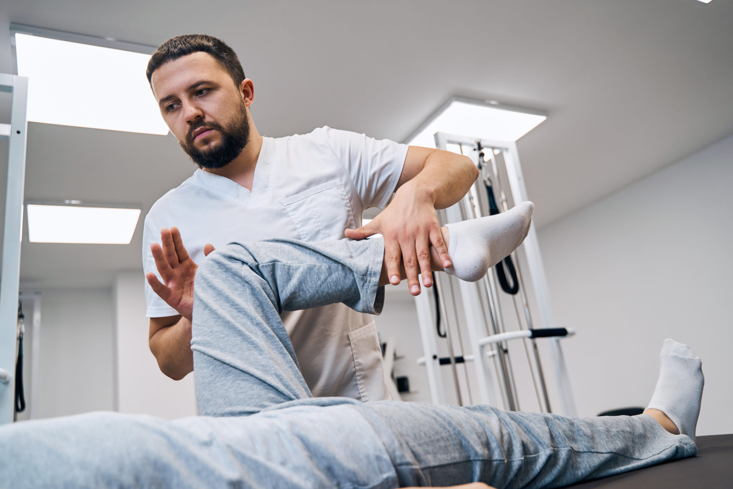
Rehabilitation following microfracture surgery.
Rehabilitation following microfracture surgery is very vital to healing. These rehabilitation protocols need to be individualized.
They are personalized depending on;
- The age of the patient
- Size of the defect in the cartilage
- Activity level
- Presence of coexisting deformities at the knee like knock knees or bowed knees e.t.c
The progression of the rehabilitation is designed based on the four biological phases of cartilage maturation which are; proliferation, transitional, remodeling and maturation.
Phases of treatment
Phase 1
Week 1 to 6 after surgery
This phase is primarily characterized by protection of the repair.
During this phase, it is imperative to control pain and reduce effusion.
Passive range of motion (ROM) should be instituted to prevent stiffness in the knee or to prevent healing with adhesions. ROM improves the nutrition of the knee which improves healing but also prevents the formation of adhesions.
Partial weight bearing with aid of crutches should be started and guided by a physiotherapist or rehabilitation specialist.
Pool or aquatic therapy can be incorporated as this creates buoyancy and therefore reduces the weight of an individual up to 25 to 50% of the weight of the individual.
Phase 2
week 6 to 12 after surgery
This phase is about restoring the individual to full weight-bearing status to resume normal activities of daily living. The individual progresses from partial-weight bearings to full weight bearings.
Activities to cater for strength training of the different muscle groups and proprioception are all instituted
Phase 3
3 to 6 months following surgery
This is in the remodeling phase of the fibrocartilage. The patient continues with the different activities that encourage range of motion and muscle strengthening.
Phase 4
The individual is able to return to full-impact activities
The outcome of microfracture surgery
Overall, the outcome following microfracture surgery is good. This study showed that children had a good clinical outcome following this surgery.
In conclusion, microfracture surgery of the cartilage improves the quality of life in patients by reducing pain. Physiotherapy after this surgery leads to a complete return to function through improving range of motion, graduation back to weight-bearing status, and muscle strengthening.
